Please Read!
As part of my tests at Stanford before my surgery, I was given two
different SPECT (single photon emission computerized tomography) scans. The scans
were used to test and measure the oxygen content of my brain and show the results in three
dimensional pictures.
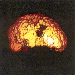
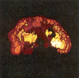
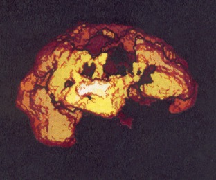
The first test was taken with me (and my brain) at rest. I was told
to relax as much as possible and try to take a nap while the test was being done.
This test was to measure the oxygen content of my brain while I was "resting".
You can see an area very well where the right side of my brain was not
getting oxygen because of the Moyamoya disease. That is the area of my brain that
controls the function of the fingers and the part of my left arm that are still numb.
That area of my brain was basically without oxygen for over five months and there
isn't much of a chance those cells will recover.
The images below reflect the results of the first test.
Scroll down slowly and keep reading!
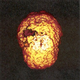
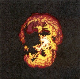
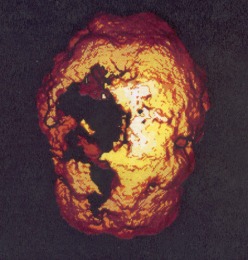
The next test was given the second day. Before this test, I was
given a shot of a medication called Diamox. Diamox is a medication which increases
the brain's ability to take in oxygen. It is commonly used in high altitude
locations to help the body absorb oxygen.
This test simulated my brain in 'action'. The pictures below
demonstrate what I've said all along.... I WAS VERY LUCKY! The affects of my stroke
could have, and probably should have been much more devastating. These pictures show
why when things started happening in a hurry while I was at work, I would loose my train
of thought.
As in the pictures above, colors are a good thing, black is bad.
Colors represent the amount of oxygen, black means no oxygen.
Where'd the right side of my brain go?
Pre-Op
Post-Op
|